

 Türkçe
  English
  Deutsch
  فارسی
فارسی 


Vaginal Ultrasonography
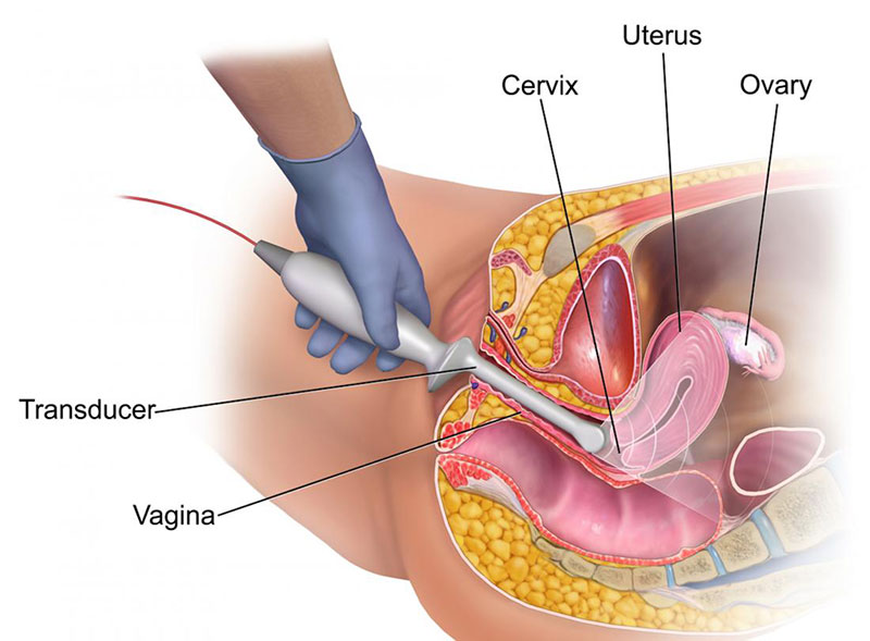
Vaginal ultrasound, also known as transvaginal ultrasound, is a non-invasive diagnostic technique that allows images to be used to evaluate organs and structures in the pelvis. This ultrasound technique; It provides rapid imaging of vaginal organs and structures, including the uterus, cervix, vagina, fallopian tubes, and ovaries.
It is the best type of test for diagnosing unexplained pain, swelling, or infections in the pelvis and for examining a growth in the pelvis. This helps determine if the growth seen is a fluid-filled cyst, solid tumor, or some other type of lump. In addition, this ultrasound technique is considered a safe procedure, although it can be a little uncomfortable.
How Does the Vaginal Ultrasound Device Work?
The vaginal ultrasound device uses a transducer that sends ultrasound waves at an inaudible frequency. The ultrasound transducer is placed on the skin and the ultrasound waves are directed towards the organs and structures within the body. Sound waves bounce off the organs like an echo and return to the transducer. The transducer processes the reflected waves and is converted into images of organs or tissues. These images are then analyzed by a computer. In addition, these sound waves travel at different speeds depending on the type of tissue encountered.
The rate at which sound waves return to the transducer and how much of the sound wave is returned are determined by the transducer as different tissue types. For best sound transmission, the transducer should be moved smoothly over the skin, while ultrasound gel should be applied to the transducer and skin to eliminate air between the skin and transducer. There are two types of ultrasound techniques and they are as follows:
Transabdominal ultrasound: In this type of ultrasound, a scan is performed by sliding it across the abdomen using transducer gel.
Transvaginal ultrasound: This type of ultrasound is performed by inserting a long, thin transducer into the vagina. In addition, the probes are covered with plastic or latex layers and are only for female use.
The type of ultrasound required will vary depending on why the ultrasound is needed in the first place. Depending on the situation, ultrasound examination is performed using one or both of these two methods.
In Which Situations Is Vaginal Ultrasound Used?
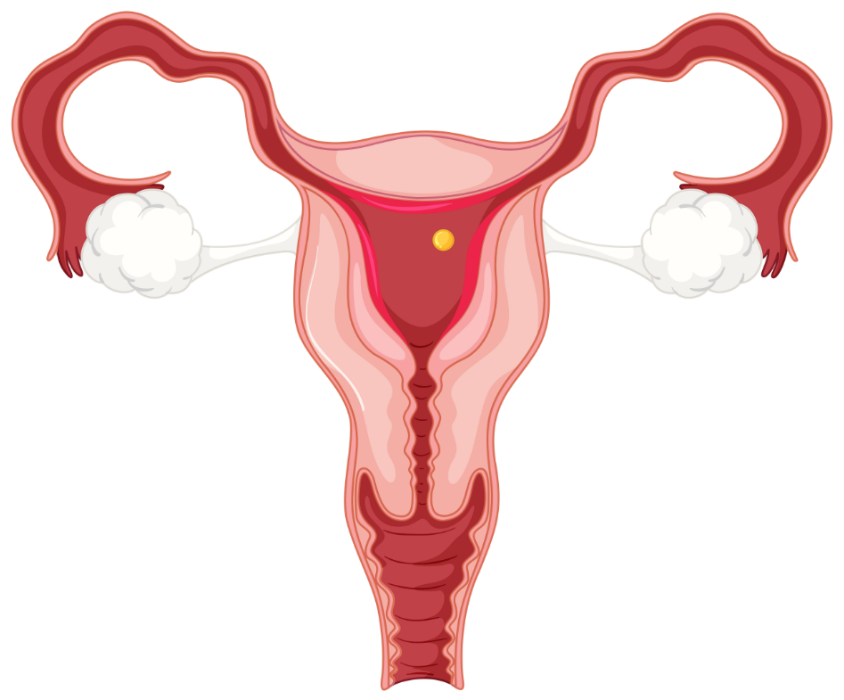
In some cases, vaginal ultrasound is needed to look at or measure the reproductive organs. Using this ultrasound, the size, shape and location of the uterus and ovaries, the density (or echogenicity) and thickness of vaginal tissues and organs, the presence of fluid or masses in the endometrium, myometrium (uterine muscle tissue), fallopian tubes or in or near the bladder, length and thickness of the cervix, changes in the shape of the bladder and blood flow from the vaginal organs are examined.
This ultrasound can provide detailed information about the size, location and structure of the vaginal masses, but is not sufficient to make a definitive diagnosis of cancer or specific disease. However, it also helps in the diagnosis and treatment of some conditions, such as:
Abnormalities in the anatomical structure of the uterus, including endometrial conditions
Benign (harmless) growths, such as fibroid tumors and cysts in the pelvis, as well as other types of tumors
Presence and location of intrauterine contraceptive device (IUD)
Pelvic inflammatory disease (PID) and other types of inflammation
Understanding the cause of bleeding after menopause
Monitoring of ovarian follicle size for infertility assessment
Aspiration of follicle fluid and egg from the ovaries for in vitro fertilization
In case of ectopic pregnancy (pregnancy that usually develops outside the uterus and occurs in the fallopian tube)
Monitoring fetal development during pregnancy
Evaluation of certain fetal conditions
How Long Is Vaginal Ultrasound During Pregnancy?
Vaginal ultrasound during pregnancy and transabdominal ultrasound are commonly used to monitor the baby's development at or before the 14th week of pregnancy. These techniques control the growth conditions of the baby, such as the baby's height, the length of the arms and legs, and the head size. It is also used to check how far the mother has progressed in her pregnancy, the position of the baby in the womb, the number of babies the mother is carrying and the amount of amniotic fluid surrounding the baby. While it can be used to look at a baby's heart, in some cases, it can act as a screening method for certain birth defects and developmental abnormalities such as Down syndrome.
Also, usually in the first weeks of pregnancy, transvaginal ultrasound is used to determine how far along the mother's pregnancy status and due date are. This method brings the probe closer to the uterus and provides a clearer visualization of the fetus during the mother's first trimester.
What Are the Risks of Vaginal Ultrasound?
In the application of vaginal ultrasound, there may be some risks depending on the person's special medical condition. Transvaginal ultrasound may cause a reaction in patients with a latex allergy because the ultrasound transducer is covered by a plastic or latex sheath. In addition, some conditions or factors such as obesity, barium remaining in the intestines of the person after the recent barium procedure, insufficient bladder filling and intestinal gas may affect the results of the test to be performed. However, vaginal ultrasound device hygiene is an important criterion to avoid a possible risk of infection.
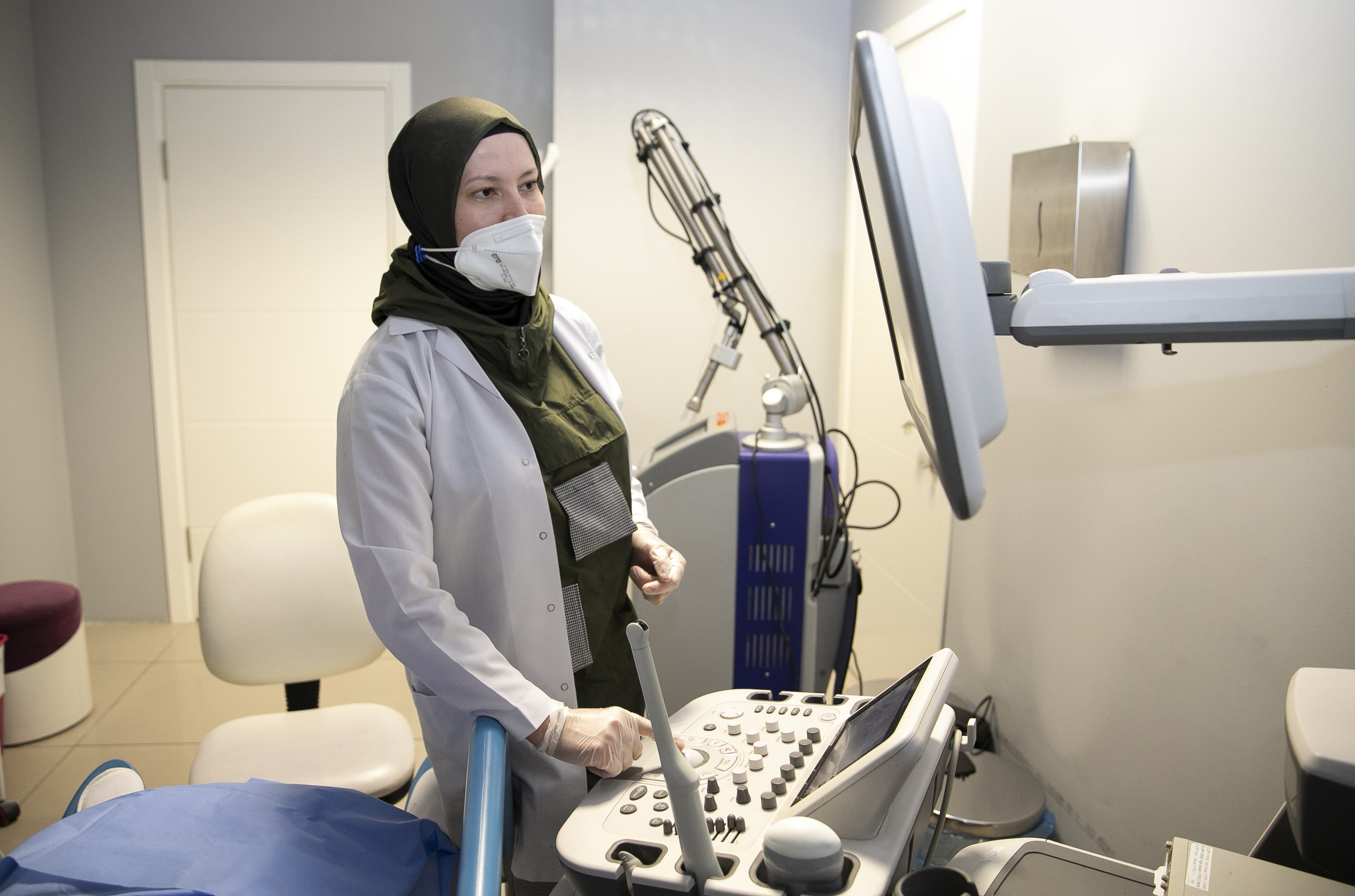
How Is Vaginal Ultrasound Performed?
In preparation for the vaginal ultrasound, if the ultrasound is not part of another procedure that requires anesthesia, fasting and anesthesia are not required before the vaginal ultrasound. In addition, at least 4-5 glasses of water should be consumed one hour before the procedure and the bladder should not be emptied until the end of the examination. For transvaginal ultrasound, the bladder should be emptied just before the procedure. Also, other special preparations may be required depending on the medical condition. Vaginal ultrasound can be performed on an outpatient basis in the office or as part of the treatment of a hospitalized patient. Procedures vary depending on the situation and hospital practices. In addition, there is no need for special care after vaginal ultrasound. Unless the person is advised differently by the doctor, he can continue his normal life immediately.